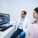
Advancing the art and science of
oral and maxillofacial radiology in Canada

3D Dental Imaging (CBCT)

The typical x-rays patients receive at their dental office are bitewings, periapical, and panoramic images. These are all two-dimensional images. Occasionally, these x-rays are insufficient to provide the required diagnostic information needed for subsequent treatment, and the dentist may require additional information in the form of three-dimensional imaging using cone beam computed tomography (CBCT).

The word “tomography” is a general term that describes any three-dimensional imaging technique that creates one or more thin section images or slices within a larger volume of anatomy that is free of overlap or superimposition from more superficial or deeper structures. Simply put, tomography, in the form of CBCT, permits a dentist to view the teeth and jaws in a clearer, more detailed manner.

Contemporary acquisition and display of tomography is now almost exclusively computer-driven, and the current standard of care for tomography in dentistry is CBCT.

Examples of common referral reasons for CBCT imaging include:

  • Suspected disease in the jaw bones.
  • Dental implant treatment planning.
  • Localization of impacted teeth.
  • Nerve localization adjacent to wisdom teeth.
  • Assessment of root canal-related problems.
  • Temporomandibular joint evaluation.
  • Jaw surgery treatment planning.

For Patients