- For Patients
For Patients
About Dental X-rays
What is a dental x-ray? Why do I need dental x-rays? Are dental x-rays safe? Other frequently asked questions. Learn more >
- For Students
For Students

Oral radiology offers diverse career possibilities in academia, research, and private practice settings. Many oral and maxillofacial radiologists combine some or all of these practice settings.
- For Dental Professionals
About Dental X-rays
27 Feb 2021 at 7:00 am

What is a dental X-ray?
X-rays are a form of radiation called electromagnetic waves. They are invisible beams of energy that pass through the body to make black and white pictures of teeth and bones.
A dental x-ray is an image that helps dentists visualize diseases of the teeth and surrounding tissue that a simple oral exam cannot see. These visualizations help with the early diagnosis and treatment of dental problems, which can help save you money, unnecessary discomfort, and maybe even your life.
Dental x-rays use one of two methods to create images. One method uses films placed in the mouth that are processed to make the picture. The second method uses plastic sensors in the mouth directly connected to a computer to create images in real-time. This second method is called digital radiography and uses much lower radiation levels than the former.
Frequently Asked Questions
Why do I need dental x-rays?
Dental x-rays (or radiographs) show your dentist or radiologist the inside of your teeth and bones. These areas are not visible in any other way. Radiographs allow early detection of oral health problems by providing information that helps diagnose dental decay, periodontal disease, and infections in your jaws. This information provides dental practitioners with the best available options for treatment.
How often do I need x-rays of my teeth and jaws?
The frequency of dental x-rays (or radiographs) is dependent on several factors, including your current oral health, your risk for dental decay and periodontal disease, and whether you have any symptoms. Your dentist considers these factors when prescribing dental x-rays. Furthermore, the American Dental Association and Food and Drug Administration have created best practices for determining the frequency of x-rays. Radiographs are recommended once or twice a year for those at higher risk for caries (cavities) or other oral disease complications.
Are dental x-rays safe?
Creating dental x-ray images uses low levels of x-radiation. The risks from these low levels of radiation exposure are relatively minimal. Nevertheless, your dentist will minimize the amount of radiation you receive using various methods, including using digital technology, customizing the x-ray machine settings to your jaw size, and protecting you with thyroid collars and lead aprons as appropriate.
What are the different types of dental x-rays?
There are two categories of dental x-rays: intraoral and extraoral. The most common intraoral radiographs are bitewing and periapical x-rays. Extraoral radiographs called Panoramic and Cone Beam CTs are less common.
Bitewing x-rays are a type of radiograph in which the film or plastic sensor has a little tab in the middle that you bite down on with your back teeth. These x-rays show details of both upper and lower teeth in one area of your mouth and are used to detect decay between tooth surfaces and changes in bone density caused by periodontal disease. Bite-wing x-ray images are also helpful in determining how well crowns fit into place whether dental fillings have been done correctly! A good bite-wing x-ray will show the tooth’s crown down to its supporting bone structure.
Periapical x-rays show the whole tooth – from the crown to beneath where it is anchored in your jaw. Each periapical radiograph only indicates two to three teeth on the upper or lower jaw. This image detects abnormalities or infections in your root structure and surrounding bone.
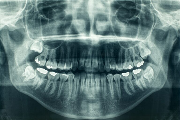
Panoramic x-rays use a machine that rotates around the outside of the head. This single x-ray image shows the structures of the entire mouth, including all teeth and jawbones and the position of fully emerged and emerging teeth. It identifies impacted teeth and aids in identifying and diagnosing tumours.
Cone Beam CT scans are similar to a medical CT scan in that it produces a three-dimensional (3D) image. The 3D imaging provides details on dental structures, soft tissues, nerve paths and bones in the craniofacial region, thereby providing greater precision in diagnosis and treatment planning of many dental and jaw diseases. A cone beam CT scan is a necessary diagnostic tool when regular dental or facial x-ray images can’t provide sufficient information.


