Exciting possibilities in clinic-based and home-based private practice are available across the country. Many OMRs combine some or all of these practice settings.
Academia:
Most Canadian and American dental schools employ an oral radiologist. The oral radiologist is typically the radiation protection officer for the institution, whose duties include establishing radiologic selection criteria, radiation safety, and quality assurance policies. Their responsibilities include the didactic and clinical education of dental, dental hygiene, and possibly graduate students. Academic oral radiologists typically participate in various university committees and conduct research.
Exciting possibilities in clinic-based and home-based private practice are available across the country. Many OMRs combine some or all of these practice settings.

Research:
Oral radiology is an integral part of general and specialized dentistry.
Areas of research include:
- Evaluation of standard imaging systems.
- The biological effects of radiation.
- The study of pathoses by radiologic imaging.
Research in oral radiology typically involves collaboration with other academic staff at dental and medical schools.
Funding for research is generally through contributions from the institution and various grants. Grant money for research is also available from private industry and organizations such as the Radiological Society of North America, which provides Scholar Grants and Seed Grants for OMRs in the early years of an academic appointment. Researchers with more experience may receive funding from the National Institute for Dental and Craniofacial Research.
The opportunities for creative activity are limited only by the imagination of the radiologist.
Private Practice + Teleradiology:
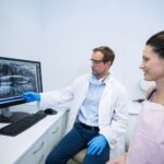
An oral radiologist may own or operate an imaging center, work with private or hospital dental clinics, or work remotely (teleradiology).
Practitioners provide radiologic reports on their interpretation of various imaging modalities, including traditional intraoral and extraoral (panoramic/skull) two-dimensional and advanced three-dimensional imaging. The most common type of three-dimensional imaging used in oral radiology is cone-beam computed tomography (CBCT). Less frequently, oral radiologists may interpret other imaging modalities, such as magnetic resonance imaging (MRI) or multi-detector computed tomography (MDCT) studies.
Examples of common referral reasons for imaging or interpretation services include:
- Suspected pathology.
- Pre-treatment implant planning.
- Localization of impacted teeth.
- Nerve/sinus association with wisdom teeth.
- Assessment of endodontic issues.
- Temporomandibular joint evaluation.
- Surgical treatment planning.